
Hip Pain in a Marathon Runner
Meet Bayleigh
Meet Bayleigh, a local personal trainer, distance runner and CrossFit athlete. Bayleigh was training for her second marathon, when following a 30km training run, she developed left hip and buttock pain. It hurt to weight-bear, and she had a visible limp when walking quickly. Bayleigh admitted increasing her running distance rapidly as her marathon was approaching and she was a little behind on her running schedule.
Following his examination, our physiotherapist ordered an X-ray which identified an acute Segond fracture at the lateral tibial plateau. This finding, along with the mechanism of injury prompted a referral to his GP who was able to order a bulk billed MRI. Unfortunately the MRI confirmed a complete ACL rupture. A referral to an orthopaedic knee surgeon was arranged. Following the orthopaedic assessment surgery was scheduled in a month later, an ACL reconstruction using a hamstring autograft combined with a lateral tenodesis.
Bayleigh presented to Total Body Physio hoping to be ready for her marathon in three weeks time.
What We Found
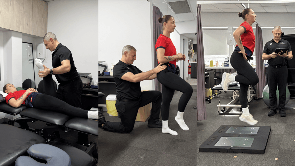
On assessment, Bayleigh presented with:
- Pain around the lateral hip and greater trochanter, with an obvious limp on faster walking
- Poor gluteal stability was noted when standing on one leg and lateral pain was present on 30-second single-leg stance test
- High levels of pain with single-leg hopping
- Discomfort across a cluster of hip provocation tests
- Pain with resisted hip flexion and on FADIR testing
- Clinical signs of generalised hypermobility (previous dancer)
Her physio liaised with her GP who organised some scans. An X-ray of her hips and pelvis was normal, however an ultrasound reported greater trochanteric bursitis.. On the surface, this pointed toward a soft-tissue problem. However, Bayleigh’s clinical examination findings did not conclusively fit the picture of a simple bursitis.
Bayleigh had pain on many of the tests that would normally be negative with a bursitis, such as the FADIR and resisted hip flexion test. Importantly, the fact that Bayleigh could not hop without significant pain was noteworthy, as bursitis rarely stops someone hopping outright. In a runner with a recent spike in training load, an inability to tolerate impact loading raised suspicion for an underlying bone stress injury, something an X-ray may not show in the early stages (Bernstein et al., 2022).
Bringing in the Sports Physician
As the clinical picture did not match a simple bursitis, and the marathon date only a few weeks away, we decided that Bayleigh should see a sports physician early rather than pressing ahead with loading or a wait and see approach, and her GP was happy to assist. This step mattered. The sports physician’s assessment agreed that the inability to hop, on a background of hypermobility and high training loads, raised genuine suspicion for a femoral neck stress fracture, and that running should stop completely until an MRI could be done.
A bursal cortisone injection was given to manage the confirmed bursitis as a holding measure, but with a clear instruction: no running until imaging had ruled out bone stress. That early referral and shared caution shaped everything that followed.
The MRI Changed the Picture
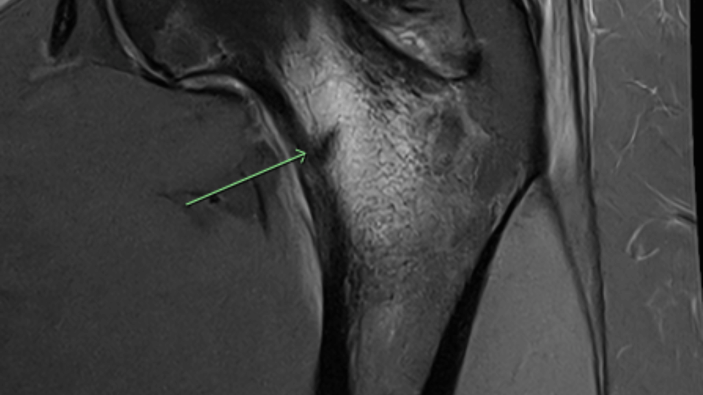
The MRI confirmed an incomplete stress fracture of the distal, posteromedial femoral neck, with surrounding bone marrow oedema. The location mattered. This was on the medial, compression side, which is generally lower risk and often managed without surgery. Tension-side fractures carry a higher risk of progressing and more often need surgical fixation (Bernstein et al., 2022).
Why Did This Happen?
Stress fractures develop when repeated loading outpaces the bone’s ability to remodel and recover. In distance runners, a rapid increase in training volume is a recognised risk factor, particularly when a large single session is added without adequate build-up (Warden et al., 2021). Bayleigh’s pain began after a sudden 30km run.
The cause was likely multi-factorial rather than a single event. A rapid spike in running volume, a background of hypermobility, biomechanics and muscle fatigue, and habitually high training loads may each have contributed (Hulme et al., 2021). Hypermobility and altered control under fatigue can change how load is distributed through the hip, which may increase stress at the femoral neck during repetitive impact. Reassuringly, Bayleigh’s bone mineral density was found to be well above average, and she had regular menses with no signs of low energy availability, which removed some of the common systemic drivers of bone stress so a conservative, staged loading pathway was then planned.
The Plan
Rehab was staged and progressed on what the hip could tolerate, not the calendar. Each phase was governed by clinical milestones and reviewed alongside her sports physician, with follow-up MRI used to confirm the bone was healing before impact was reintroduced.
Phase 1: Offload and protect
Swelling control, quad activation, gentle range of motion targeting full extension, and gait retraining with crutches. The graft is most vulnerable here — early care is about protecting it while beginning to move.
Goals: settle irritability, protect the healing bone, and maintain fitness without impact.
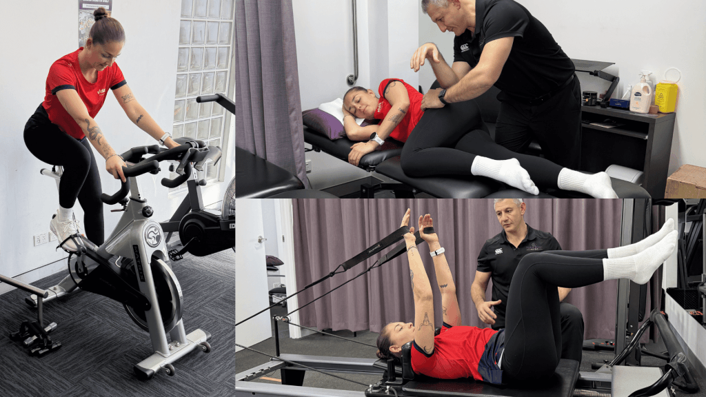
The early priority was to take load off the femoral neck while keeping the rest of the body working. Bayleigh used crutches initially, then a period of reduced weight-bearing, progressing as pain settled. Load was kept symptom-free, since a symptom-driven approach that avoids pain during, after, and the day following loading is central to managing low-risk bone stress injuries (Warden et al., 2021). Rehab included:
- Non-impact conditioning on the bike, and pool work for cardiovascular fitness
- Soft tissue work and dry needling to manage irritability and muscle tightness/overactivity
- Deep core activation exercises progressing onto the Pilates reformer
- Gentle isometric and low-load activation exercises targeting her glutes, adductors and lower limb, kept at/below low pain threshold.
- Education that the goal was to let the bone recover
Unfortunately, an early mild flare up after a period of increased load due to working as a personal trainer combined with incidental walking was a useful reminder that the posterior cortex was slow to settle, and that patience mattered.
Phase 2: Rebuild strength, stay impact-free
Goals: restore lower-limb strength and load capacity while keeping impact out until the bone allowed it.
With bone marrow oedema markedly improved on repeat MRI at the 12 week mark and a normal clinical exam, including pain-free hop testing, Bayleigh was cleared to upgrade her strengthening while still holding off running for a further period.
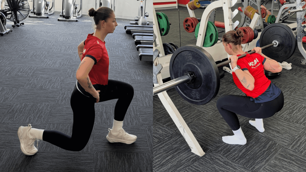
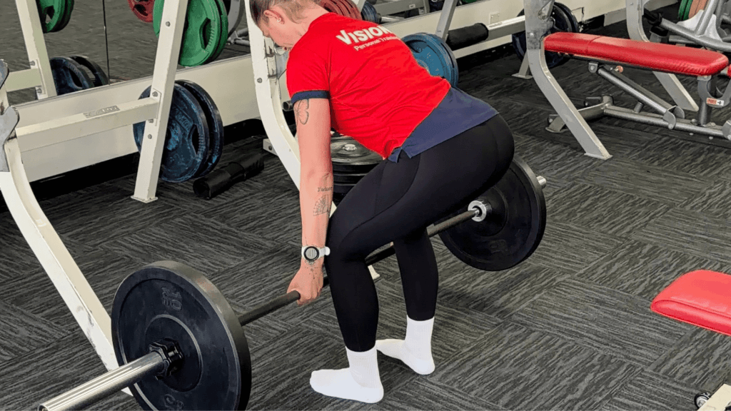
- Progressive gym-based loading: barbell squats and deadlifts, machine work including leg extension, seated hamstring curl, soleus and calf work,
- Single-leg strength and control: single-leg squats, lunges, and hip thrust progressions
- A staged introduction of low-level plyometrics once strength and control allowed
- Ongoing objective monitoring, with an in-person review and hop testing used to confirm she was ready before advancing
Building both local hip and proximal strength before reintroducing running is consistently recommended following bone stress injury (George et al., 2024). The rule throughout this phase was simple. If pain returned, back off, let it settle, and rebuild.
Phase 3: Return to running
Goals: reintroduce impact, restore running capacity, and address the biomechanics that contributed in the first place.
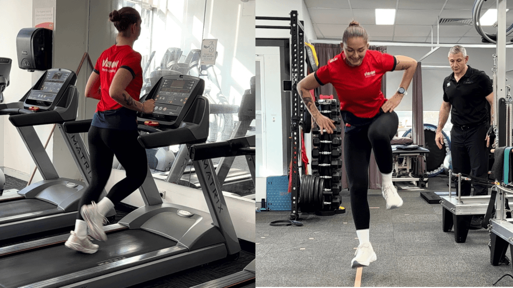
Once strength, single-leg control, and pain-free hopping were established, running was reintroduced in a graded way. Resolution of bony tenderness, pain-free walking, and passing strength, functional and loading tests are the components recommended before reintroducing running (George et al., 2024).
- A structured walk-jog progression, building total volume gradually over weeks before increasing continuous running
- Continued strength and plyometric development to support running load: broad jumps, straight-leg bounds, drop jumps, and single-leg jump variations
- Attention to running mechanics and load management
- Clear self-management strategies to recognise early warning signs and adjust load before problems developed
Objective Outcomes
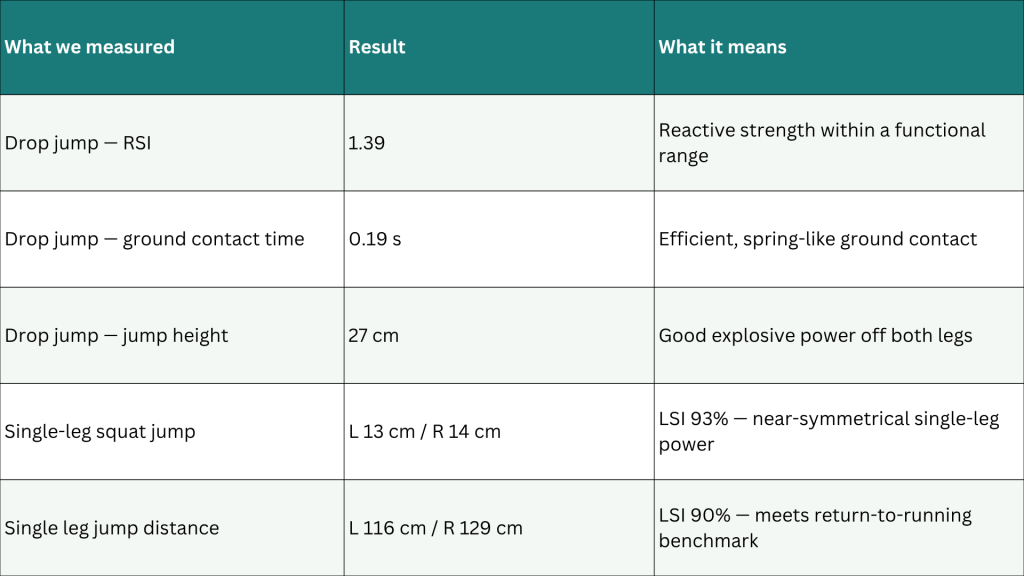
Rehabilitation was progressed using objective markers, not time or pain alone (Hoenig et al., 2023). Alongside clinical milestones, jump and force-plate testing was used. At her return-to-running testing, the injured left side sat within accepted symmetry thresholds across reactive, power and single-leg measures, which supported a graded return to running.
The jump testing showed that Bayleigh’s reactive strength and vertical output had recovered well, with only a small residual difference in single-leg jump distance, all within the symmetry range used to support a return to running.
Where Is She Now?
Bayleigh is happily back running and training normally. Since recovering she has completed two half marathons and a HYROX event. Recent testing on our VALD system tells a reassuring story. On the previously injured left side she now tests stronger than the right and she had 99% symmetry on jump testing. Her reactive hop scores also sit well within normal ranges. Her numbers reflect what would strive for following a comprehensive rehabilitation program.

Key Clinical Takeaways
- A hip or buttock “bursitis” that stops an athlete hopping deserves a second look. When impact loading is disproportionately painful, consider an underlying bone stress injury, even when early X-ray and ultrasound point elsewhere (Hadjispyrou et al., 2023).
- Plain X-ray may miss a femoral neck stress fracture early. Where suspicion is high, MRI provides what plain film cannot.
- The location guides management. Compression-side (medial) fractures are often managed non-operatively, tension-side fractures more often need surgical fixation (Bernstein et al., 2022).
- Early referral and shared decision-making can change the course of an injury. Stopping running before imaging may have prevented a low-risk fracture from progressing.
- Return to running is guided by bone healing, strength and objective testing, not time or pain resolution alone. Pain settling is not a proxy for readiness (George et al., 2024).
If you or someone you care for has an injury, a flare up, requires some rehabilitation or experiences an increase in pain, give the clinic a call on 9713 2455 or book online.
Check out our socials for videos on exercises and tips!
Share to
Facebook
LinkedIn
Pinterest
X
Email