
Same Injury, Different Foot: When a Lisfranc Injury Needs Surgery
Meet Kate
Kate is a 40-year-old recreational netballer who enjoys playing basketball with her son and going for walks. During a weekend netball game, her right foot rolled inward through the arch and she heard an audible pop. There was no big collision or dramatic fall, but pain followed and walking became difficult.
Kate felt like she had been here before. She had previously sustained a Lisfranc injury to her left foot, which recovered exceptionally well with conservative management and rehab. Kate initially hoped this new injury would be easy to manage with some physio also. However, her difficulty weight-bearing and the severity of her symptoms raised concerns that something more significant had occurred.

What We Found
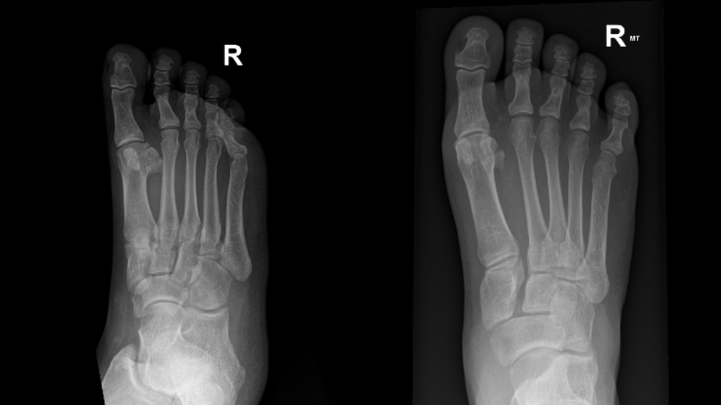
An X-ray was first requested to rule out a navicular fracture and assess the Lisfranc joint. It showed widening at the Lisfranc joint, several densities suspicious for small bony fragments and swelling over the top of the foot. The radiologist recommended further imaging.
Within days, Kate was reviewed by an orthopaedic foot and ankle surgeon. Assessment showed:
- Displacement and widening through the Lisfranc and midfoot joints on X-ray, along with suspected small bony fragments, compared with normal alignment on a comparison X-ray of the left foot, where she had previously been injured.
- Bruising on the sole of the foot (plantar ecchymosis), a key warning sign for Lisfranc injury.
- Midfoot swelling, though not severe enough to delay surgery that was likely required.
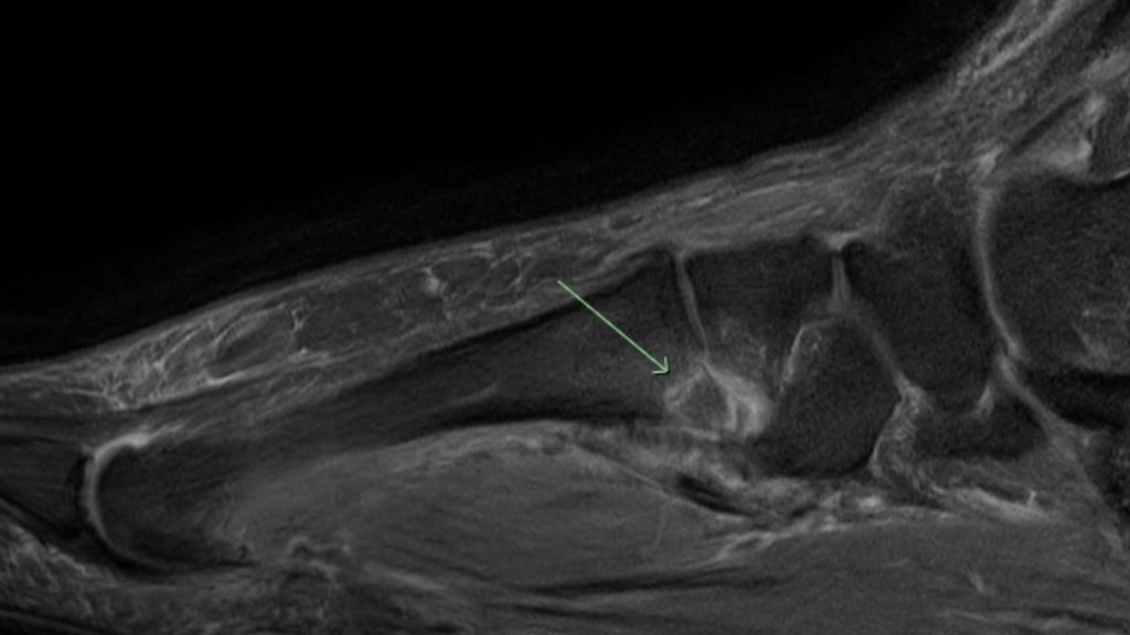
MRI confirmed an unstable, displaced Lisfranc fracture-dislocation with disruption of the supporting ligaments.
Why Does Early Imaging Matter?
Lisfranc injuries are frequently missed on plain X-ray because the signs can be subtle. Plantar bruising and midfoot pain after a twisting injury warrant a high index of suspicion and consideration of early advanced imaging (McDermott et al., 2024).
When clinical suspicion remains high, further investigation may be necessary:
- CT scan: provides detailed assessment of the bony architecture and helps identify small fractures not seen on X-ray.
- MRI: helps identify ligament disruption and associated soft tissue injury (Tang et al., 2024).
- Weight-bearing imaging: may reveal instability that is less apparent when the foot is unloaded, provided
weight-bearing is clinically safe. Weight-bearing CT scans show promising results for subtle injuries (Poulsen et al., 2025).
In Kate’s case, X-ray and MRI findings established the extent of the injury and helped guide the surgical decision.
Understanding Lisfranc Injuries
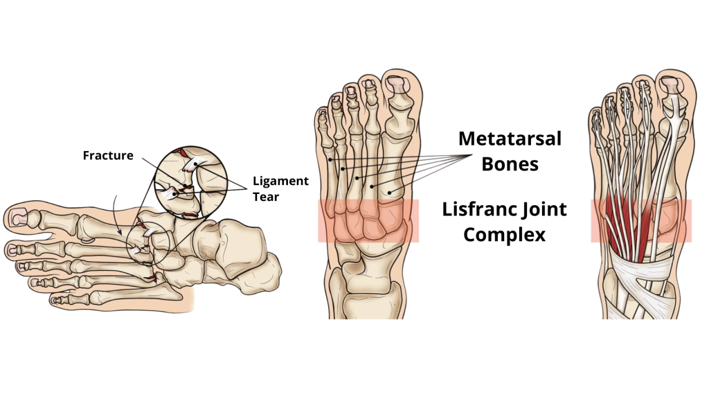
The Lisfranc complex is the group of joints and ligaments where the long bones of the forefoot (metatarsals) meet the smaller midfoot bones. The Lisfranc ligament anchors the base of the second metatarsal to the medial cuneiform, helping keep the midfoot stable as we push off (Chen et al., 2020).
Think of the midfoot as a stone archway. The bones form the structure, while the ligaments help hold the arch together. If the supporting ligaments fail and the bones shift out of position, the arch can lose its stability.
Lisfranc injuries range from mild sprains to complex fracture-dislocations. Injuries often occur when load is driven through the foot with the toes pointed and the midfoot twisted. They can occur during sports like netball, football, rugby and other activities involving pivoting, landing or twisting through a loaded foot, in addition to falls and crush injuries.
Management
The key factor when it comes to management is not simply whether a Lisfranc injury is present, but whether the midfoot remains stable.
A stable Lisfranc injury means that the midfoot joints remain correctly aligned under load. An unstable injury means that the joints may separate or shift. Importantly, an injury can appear non-displaced on an initial scan yet still be unstable when weight is applied, so “non-displaced” does not necessarily mean “stable”.
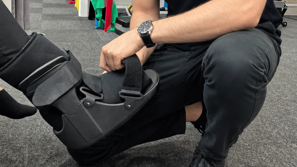
- Stable, non-displaced injuries can be managed with approximately six weeks non-weight-bearing in a cast or CAM boot, followed by graduated loading and rehabilitation.
- Unstable injuries, with or without obvious displacement, generally require surgery. Options include open reduction and internal fixation (ORIF) with plates and screws, or a technique called “primary arthrodesis” where selected joints are fused. The choice depends on the injury pattern, joint damage and the surgeon’s assessment (Poutoglidou et al., 2024).
Without appropriate management, an unstable Lisfranc injury may lead to persistent pain, progressive deformity, impaired walking mechanics and post-traumatic arthritis.
The Surgical Decision
This was the defining difference in Kate’s case. Her previous left-sided injury healed successfully with conservative management, protection and rehabilitation as she had a stable Lisfranc injury. Her right-sided injury, however, involved displacement of the midfoot joints and disruption of the supporting ligaments. Although both injuries involved the Lisfranc complex, they were not mechanically equivalent. A boot can protect a stable midfoot, but it cannot pull displaced joints back into alignment. If left unstable, these injuries may lead to progressive arch collapse, chronic pain, stiffness and post-traumatic arthritis (Sain et al., 2023).
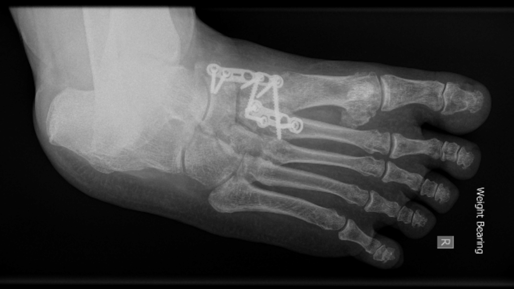
Due to these factors, the surgeon advised that the operative approach was the necessary pathway for Kate, and she had surgery within a week of injury. The displaced midfoot joints were realigned and stabilised with plates and screws (ORIF). ORIF aims to restore alignment and hold the injured structures in position while they heal, preserving the joints rather than deliberately fusing them.
Her postoperative plan involved two weeks in a plaster cast for wound healing, followed by a CAM boot with arch support. She was to remain non-weight-bearing for approximately six weeks in total. Hardware removal was planned for around six months, following ligament healing.
Surgery is not the answer for every Lisfranc injury. The decision depends on midfoot stability, displacement and structural damage. For Kate, whose injury involved a displaced and unstable fracture-dislocation, the surgeon recommended fixation to restore a stable, functional foot.
The Role of Physiotherapy
Surgery restores alignment and stability, but returning to comfortable walking, exercise, sport and normal tasks of daily living requires time and structured rehabilitation. For Kate, the long-term goal is not simply to heal the injury. It is to regain confidence in her foot and return to the activities that matter to her, from everyday family walks to playing basketball with her son, domestic activities and recreational netball.
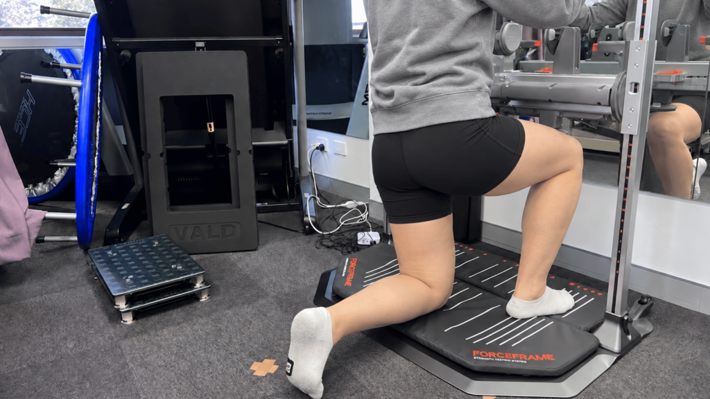
Kate is currently undergoing rehabilitation. Her rehabilitation follows a criteria-based pathway and progresses according to tissue healing, surgical restrictions, symptoms and functional milestones rather than a fixed timeline alone. In our clinic, we track this progress using objective measures of movement, strength, balance and functional capacity, including VALD strength testing where appropriate.
Stage 1: Protect and restore comfortable movement
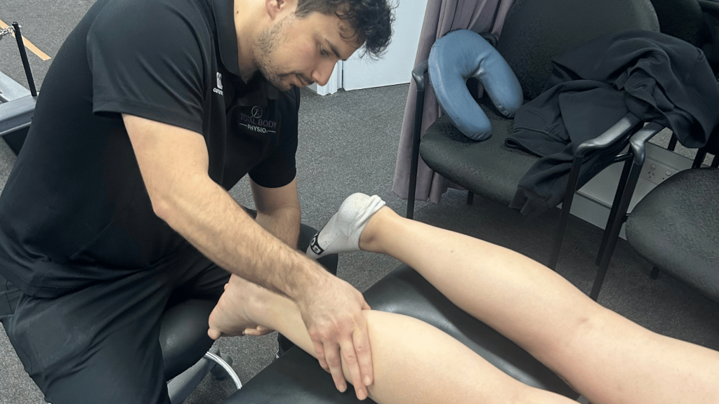
Early physiotherapy focuses on:
- Swelling management.
- Soft tissue desensitisation.
- Mobility of the extensor tendons over the top of the foot.
- Restoring ankle movement within the surgeon’s restrictions.
As healing progresses and increased loading is allowed, weight-bearing can gradually build in the boot before transitioning into supportive shoes.
Stage 2: Rebuild walking and everyday function
Once cleared by the treating surgeon, rehabilitation can progress towards:
- Graduated weight-bearing in the prescribed boot.
- Gait retraining and restoration of walking mechanics.
- Improving ankle mobility within the permitted range.
- Progressive calf and lower-limb strengthening.
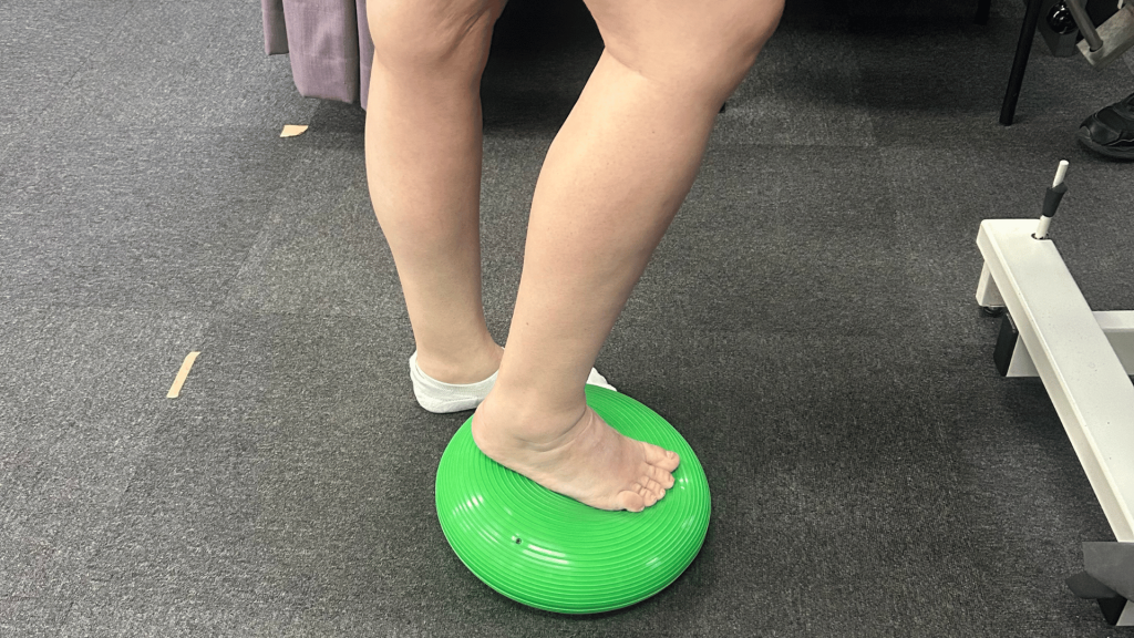
- Balance and proprioceptive exercises when appropriate.
Early functional goals may include managing household activities, walking short distances comfortably and gradually returning to family outings. Walking volume should increase in response to a reduction in symptoms and swelling, an improvement in movement quality and the treating team’s guidance.
Stage 3: Restore strength and capacity
The priority is to prepare the foot and lower limb for increasing physical demands.
As healing progresses, rehabilitation can incorporate:
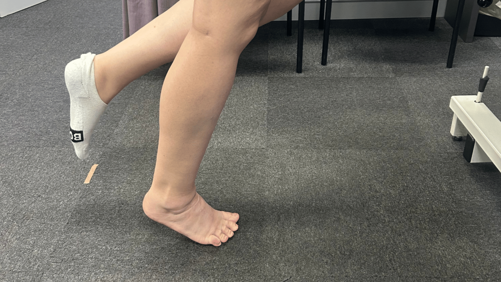
- Progressive calf raises, advancing from supported bilateral exercises towards unilateral loading.
- Resistance-based strengthening for the lower limb.
- Balance and single-leg control.
- Progressive gym-based rehabilitation.
- Gradual restoration of push-off strength and walking endurance.
The focus is on rebuilding the strength, control and capacity needed for longer walks, gym training and more demanding daily activities. Any planned hardware removal may require reassessment and temporary modification of loading, depending on the procedure and surgeon’s advice.
Stage 4: Return to running and sport (as determined by the patient’s goals)
The aim is to restore the capacity to run, land, accelerate and change direction safely.
If returning to netball is appropriate, later-stage rehabilitation may include:
- Progressive impact loading and jogging.
- Plyometric drills including hopping and landing.
- Acceleration, deceleration and change-of-direction training.
- Sport-specific conditioning.
- Functional testing to assess strength, balance, movement quality and tolerance to impact. In our clinic, we use VALD strength testing equipment to assess various markers at this stage.
Progression should be guided by symptoms, functional performance and specialist clearance, with side-to-side comparisons used where appropriate.
A return to netball following a displaced Lisfranc fracture-dislocation is a substantial rehabilitation goal. Recovery often takes many months, and timelines vary according to the injury, surgery and individual progress.
KEY TAKEAWAYS
- Not all injuries involve a fall, a collision or a dramatic accident. A “minor” twist through the arch with a pop and plantar bruising may indicate a Lisfranc injury. Early imaging matters.
- X-ray may raise suspicion, but MRI or CT is often needed to confirm the extent of bone and ligament damage. A comparison view of the other foot can help.
- Stability dictates management. Stable injuries may recover well without surgery, while displaced or unstable injuries usually require surgical stabilisation (Grewal et al., 2020).